Nerves of an Escape Artist
By Robert Frederick
Studying the neurons of a most elusive animal required a new trap, which worked—at least for a little while.
Studying the neurons of a most elusive animal required a new trap, which worked—at least for a little while.

If only hydrae weren’t so fragile: They are ageless, have a nervous system that is constantly in flux, and so other-wise would be excellent model organisms for neuroscientists to study for the relationship between neural activity and behavior. But even the gentle microfluidic devices developed to immobilize tiny, transparent roundworms C. elegans beneath a microscope rip hydrae apart. “We had to redesign everything,” says Jacob Robinson, a neuroengineer at Rice University who developed a new device to study the delicate creatures.
Krishna N. Badhiwala, Jacob T. Robinson, et al.
Ripped-apart hydrae are quite interesting to researchers, too, particularly because they regenerate. Some researchers have even put hydrae into a blender, reaggregated their cells in a centrifuge and found that hydrae still re-form. “As the animal is damaged it repairs itself,” Robinson says, “The brain is constantly changing and rewiring as new neurons are formed.”
It’s those tantalizing features of this cousin of the jellyfish that neuroscientists are keen to study, but there’s a limit to what researchers can learn from ripped-apart hydrae, no matter how carefully they’re dissected. Indeed, compared to hydrae, neurological connections in other model organisms, such as C. elegans, are “relatively static,” Robinson says, so “the overall connectivity between all of the cells is relatively the same across different animals throughout their lifetimes.” Among hydrae in the same species, the number of neurons can change dramatically—by factors of 10.
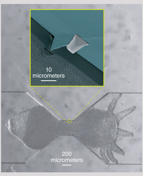
One of the microfluidic devices Robinson and his team developed to study these dynamic creatures pushes a hydra into an hourglass-shaped container. The container is embedded with a tiny electrode that the team had previously developed to study the electrophysiology of C. elegans.
Krishna N. Badhiwala, Jacob T. Robinson, et al.
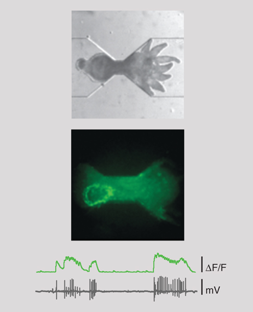
In a proof-of-principle study published in the Royal Society of Chemistry’s journal Lab on a Chip, the team demonstrated the capacity to capture the animal’s electrophysiology and neural activity simultaneously, allowing researchers to study the “activity of muscle cells and the group of neurons responsible for motor function during body column longitudinal contractions,” the team writes.
Essentially, those “body column longitudinal contractions” enable inchworm-type locomotion that allowed the hydrae to escape. It took hours. But prior to the work of Robinson’s team, typical recording periods lasted only a few minutes.
With hours to work with, the team also showed that they could clearly image all the neurons in a living hydra that had been genetically modified with green fluorescent protein (below).
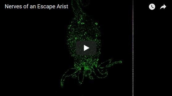
Video by Krishna N. Badhiwala, Jacob T. Robinson, et al.
Robinson says, “What’s interesting about the structure of this nervous system is that the neurons are distributed almost uniformly throughout the whole animal.”
The overall goal is to understand the relationship between “what the animal is doing and what the animal is thinking,” and that is just getting started, Robinson says. But given that the common ancestor between hydrae and humans is so ancient that it could be the first creature with a nervous system, a better understanding of a living hydrae’s nervous system should provide valuable insights into how our own nervous system evolved.
An audio interview with the researcher, Jacob Robinson:
Click "American Scientist" to access home page

American Scientist Comments and Discussion
To discuss our articles or comment on them, please share them and tag American Scientist on social media platforms. Here are links to our profiles on Twitter, Facebook, and LinkedIn.
If we re-share your post, we will moderate comments/discussion following our comments policy.