The Changing World of Microscopy
By Fenella Saunders
Plant cell biologist Harry (Jack) Horner has seen microscope imaging technology change a lot over his career, but the field isn't fading out as some had predicted.
December 20, 2022
From The Staff Biology Technology Botany
Now an emeritus professor at Iowa State University, Harry (Jack) Horner's research encompassed several diverse areas in development and cell biology, often with a focus on plants, with both theoretical and practical importance. While a graduate student at Northwestern University, Horner received a Sigma Xi GIAR grant, which helped him buy equipment as he started getting into electron microscopy. He would go on to a postdoc at Iowa State University, where he would become director of the university’s Microscopy and NanoImaging Facility. Horner spoke with American Scientist’s editor-in-chief Fenella Saunders about how microscopy has changed—and is still changing—along with changes to the scientific research enterprise, as well as how he stays active in research today. This interview has been edited for length and clarity.
Your career in microscopy involves both engineering and biology. How did that come about?
I started out at the Colorado School of Mines, in Golden, for two years. I decided that engineering was not for me, so I transferred to Northwestern University, north of Chicago. I was an undergraduate biology major without really any decision as to what I wanted to do.
When I got into my fifth year, actually—I took an extra year—I decided maybe I wanted to teach. I took the minimum number of education courses to teach, and when I was near the end of my fifth year, I student taught. I went out and spent the whole weekday teaching. At that time, I was also doing an independent research project with a faculty member in the biological sciences department. I enjoyed the research, and I truly enjoyed the teaching. At that point I said to myself, where can I do both?
That of course pushed me to start my graduate degree. I did a master’s in one year and a PhD in four years. During that time Sigma Xi was kind enough to give me a grant to purchase a diamond knife. I had just started in electron microscopy. I went from the Evanston campus downtown to the medical school, where the head of the anatomy department said, “Come on down and use our microscopes. We don’t know anything about plants, and we’d love to learn.” That was a wonderful introduction during the last year of my graduate work, and that’s what pushed me into finding a postdoc in which I’d continue doing electron microscopy. I applied for an NIH postdoctoral fellowship at the end of my PhD and received a two-year postdoctoral fellowship. I went to Iowa State University without any intention of staying. But Iowa State came and asked me if I was willing to stay. They would create a position and match all the other offers. I liked Iowa State. I liked the Midwest. So I accepted the position in 1966.
Four years later, I began directing a microscopy center on the campus. I continued to direct that for 47 years. I was involved in cell biology in terms of my research and my graduate students. I also served on many PhD and master’s committees during my time there. It was a wonderful experience.
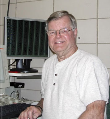
Courtesy of Harry Horner
How would you describe your research area?
I’ve been involved in a variety of research areas, partly because of funding and partly because of my own specific interest. I’ve worked on microsporogenesis and megasporogenesis, bacterial nodulation in higher groups of plants. I’ve worked on nectarines, male sterility in plants, and several other areas. So I’ve had a broad interest in a variety of research areas, which fortunately didn’t penalize me like many of the researchers who’ve been focused in one research area most of their careers. I had the fortune of being able to spread my interests and work with a number of researchers, not only at Iowa State, but in other parts of the world: Germany, Mexico, and Belgium.
Tell me more about your research in male sterility in plants.
I worked with several other researchers who carried out molecular and metabolic studies. My particular area was following the actual development of the anthers with light and electron microscopy. We meshed our specific areas to study the basis for male sterility in a variety of both crop and non-crop plants.
So with anthers we studied how they were initiated in the flowers and followed the development of the male cells in what's called the nurse cell, the tapetum. We followed how they developed, when they aborted, and other aspects of the research, to determine the actual control based on the genetics of the systems.
It really evolved into trying to develop male sterile soybeans, which hasn’t really occurred completely yet. A colleague of mine who was in the agronomy department—a world-renowned geneticist—we were trying to develop a male sterile line that was environmentally stable in order to enact a hybridization. You could have offspring that had the compatible characteristics in the environment in which they were growing. We had support from Pioneer here in Iowa, and we even got a patent on one of the lines we were working on. Unfortunately, my colleague passed away, and that ended that aspect of that research.
Overall, it sounds like you didn’t quite get away from your engineering roots. Were you interested in the imaging machinery aspects of it?
Yes, in a way. I learned one thing that I always tell my students, and that is, even if you switch areas, what you learn in one area quite often carries over into another area. Obviously some of the engineering I picked up over those two years became quite applicable to what I was doing in running the microscopy center. I taught graduate level courses in microscopy for about 40 years. The engineering did help in many ways.
But back in the mid-1980s, I had a dean tell me that microscopy was going to die because molecular biology was going to take over everything. However, that hasn’t occurred, and I don’t think it ever will occur. Microscopy is still a very important area to complement other areas of research. And it’s still changing. There’s just a lot of innovation that’s going on.
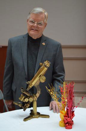
Courtesy of Iowa State University
You retired in 2018, but you still have papers coming out, including a highly cited article about calcium oxalate from 2020. Are you still doing collaborative work?
I’m not doing any more collaborative work, but I am trying to keep active with mini-reviews. It keeps me busy and out of trouble.
Calcium oxalate is a product in animals. It’s typically a pathological condition that forms kidney and bladder stones. That’s a big problem area for both humans and animals. But in plants, calcium oxalate is not a pathological condition. It’s a normal product in many plants that has a variety of functions. There’s no one function for that crystallization in plants. It can be sequestering calcium or oxalate. It can be for light gathering and reflection. There are other functions that are shown. Stinging nettle plants have crystals at their tips. That’s a protective function. But It’s still one of the challenges for plant-oriented people to figure it all out.
My first interest in calcium oxalate, really, was when we were looking at bacterial leaf nodules, which occur in two families of plants. We were looking at the nodules, and it turns out there were crystal cells, because the calcium oxalate forms in living cells around the bacterial nodules. It turned out that the sections I cut showed a variety of developmental stages and characteristics within the cell structure that had never been published on before. So that’s what got me started on calcium oxalate crystals in plants.
Unfortunately, I never was able to get any grant support for my research. I think I published 50 papers in that area with no support whatsoever. But I was able to do the new research and get it published.
Getting reproducible results in microscopy is important. But is it a skill that anyone can learn, or is it as much an art as a science with sample preparation and getting everything to work right?
All the years I’ve taught the three different levels of microscopy—light microscopy, scanning electron microscopy, and transmission electron microscopy—there are students who pick it up naturally, like learning how to play a piano or some other musical instrument, and there are other ones who just don’t have the aptitude for it. They have a very difficult time, and they generally don’t continue on in the area of microscopy.
So at the facility that I directed, on a yearly basis, we handled more than 100 researchers, both on campus and off campus, and many of them did not want to do the microscopy work. They wanted to have the work done. So we charged for that. But there were researchers who wanted to learn the different techniques for imaging, and that’s why we had the courses to train them.
Besides the difficulties in doing microscopy for themselves, have the kinds of obstacles that researchers encounter changed over the years?
I think it’s very difficult for a faculty member who’s doing research to continue on with a viable research program without getting funding, whether from NIH, USDA, or NSF. If they don’t get that funding, I’ve had a number of colleagues retire even with, in my opinion, very fine research interests. So that has changed. And of course then the major granting institutions have special focus areas. If you don’t fall within them, then the chances of you getting any amount of funding are very difficult.
What are things that scientists who are coming up right now might do to have a better experience, or that the research enterprise might change to improve?
In today’s atmosphere, it’s very difficult for any researcher, whether young or in the middle stages of their life, to exist without being able to get funding. But I think the process of a beginning graduate student hasn’t changed. They look for a professor who they want to work with and who has a research area that they’re interested in. When I started with my major professor, I could choose whatever I wanted to do in research, which is not really possible today. Today, the major professor is being funded for a specific area of research, so the student has to pick some aspect of that. When they finish, of course, and they’re getting a job—whether in academia or private industry—in part it’s based on their own individual interests and in part on job availability. Students getting their PhD today typically end up going on postdoctoral fellowships, and those postdoctoral fellowships can extend way longer than one or two years. I’ve seen some students going for six or seven years. To me that’s way too long. But if they can’t find a job, then staying on in a postdoctoral fellowship is the viable way to go.
As far as advice, I think a student needs to know that they’re in charge of their own destiny. They’re the ones who can decide with whom they want to work. If they’re unhappy with whomever they’re working with or with a research project, it’s up to them to make the change. Sometimes that’s possible and sometimes it isn’t. But knowing that they have the freedom to make those decisions is important for their lives. So that would be my recommendation: Stand up for what you want and what you believe in.
American Scientist Comments and Discussion
To discuss our articles or comment on them, please share them and tag American Scientist on social media platforms. Here are links to our profiles on Twitter, Facebook, and LinkedIn.
If we re-share your post, we will moderate comments/discussion following our comments policy.