Up Close with a Squash Bug
By Felice Frankel
Maria Eisner explains how she uses electron microscopy to take a closer look at insects
Maria Eisner explains how she uses electron microscopy to take a closer look at insects

DOI: 10.1511/2005.52.166
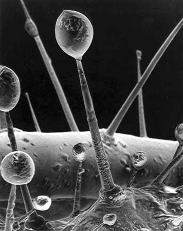
Do you know how it is when you see an image of some sort and it becomes burned into your synapses? (I hope my neuroscientist friends forgive my scientifically imprecise language.) Well, it happened to me when I saw one of the covers of Science in July 1998. I was stunned by its drama and content. A scanning electron micrograph of the surface of the pupa of the ordinary squash bug revealed a startling armature of glandular hairs that exude an oily chemical cocktail, seen forming droplets at the tips. Maria Eisner is a research assistant at Cornell University. Her photographs are featured in For Love of Insects (Harvard University Press, 2003), by her husband and long-time collaborator, Thomas Eisner.
Reprinted by permission of the American Association for the Advancement of Science.
F. F. Your material is on a scale within the range of optical microscopy. Why did you decide to use electron microscopy to capture the image?
M. E. With electron microscopy you can work at a much higher magnification, and you have more depth of field. The pictures almost seem to be three-dimensional.
F. F. It appears that you had no problem with electron-beam damage. How can that be with what seems to be a very delicate sample?
M. E. The droplets at the tips of the hairs consist of a viscous secretion that is not very volatile and therefore can stand up under the electron beam at high vacuum.
F. F. Your image is captured on film, but most scanning-electron-microscopy images these days are captured as digital files. Do you think there is a difference?
M. E. Our scanning electron microscope is an old instrument that is not connected to a computer, so we have no way of comparing photographic and digital images.
F. F. Can you explain to our readers how you prepare your samples?
M. E. The most important thing about an item to be viewed under an SEM is that it be completely dry, inside and out. Yet most biological specimens, like pieces of plants or a caterpillar, contain water. Just letting them dry by air causes them to lose their shape, in most cases. In order to avoid such distortions I let my specimen sit in 70-percent alcohol for some hours or a day and then change it to increasingly higher concentrations of alcohol, until I end up with 100 percent. Now I can use a small apparatus, a critical-point dryer, in which the specimen is dried under special conditions of pressure and heat, that leave it natural-looking, mostly without distortions. After I glue it to a microscope stub, the specimen is subjected to a vacuum in yet another small apparatus and "sputtered" with gold atoms. The covering layer of metal keeps my specimen from charging and from burning up while it is exposed to the electron beam in the microscope.
Another way of dehydrating specimens is by freeze-drying. The pupa of the squash beetle that I used for this image was frozen on the stage of the freeze dryer and dried for several hours under those conditions.
F. F. A number of SEM images are digitally colored these days. In fact, I add color myself, but of course I always indicate that I’ve done so in the process. Do you have any opinions about digitally coloring SEMs?
M. E. We have never done it nor tried to do it, because we are happy with our black-and-white images. Coloring them is to us like playing around with the images to achieve special unnatural effects.
Click "American Scientist" to access home page
American Scientist Comments and Discussion
To discuss our articles or comment on them, please share them and tag American Scientist on social media platforms. Here are links to our profiles on Twitter, Facebook, and LinkedIn.
If we re-share your post, we will moderate comments/discussion following our comments policy.