The Nets Around the Neurons
By Sandra J. Ackerman
In a mass of tissue as densely packed and hard-working as the brain, even the holes among the structural elements have jobs to do.
In a mass of tissue as densely packed and hard-working as the brain, even the holes among the structural elements have jobs to do.

DOI: 10.1511/2016.118.6
We’re accustomed to thinking of the brain as an organ that not only generates our mental constructs but retains them as well. “Hold that thought,” we’ll say. Of someone who never seems to forget anything, we might exclaim, “He has a mind like a steel trap!” How the brain creates a memory, stores it among numerous strengthened synapses that constitute a particular signaling pathway, and is then able to retrieve that memory after a lapse of days or decades, is something that neuroscientists are just beginning to understand. We don’t consider it remarkable that a nonagenarian, for example, can recall with perfect clarity how he played stickball in the city streets 80 years ago or that a retired professor can tell you the name of her second-grade teacher; yet these feats of memory are difficult to explain.
Of course the neurons, with their signal-transmitting synapses, play a leading role in the explanation, as do the axons (the extended branches of a neuron that emit the signals) and the dendrites (the spiny protuberances of neurons that receive them). This much has been known for some time. Lately, however, some non-neural elements of the brain have been receiving their share of attention, such as the variety of proteins and sugars that make up the extracellular matrix. As is so often the case in living systems, the more closely each element is examined, the more complexity it reveals.
In recent studies and in presentations at the 2015 annual meeting of the Society for Neuroscience, several research teams have laid out evidence that the brain’s extracellular matrix—far from being an inert substance that serves only to hold the busy neurons in place, as was previously thought—acts dynamically to take part in several major brain functions, from development to maintaining the stability of synaptic connections.
Even within the obscure world of the extracellular matrix, one of the least well known elements has been the perineuronal net, a structure that seems almost to have a penchant for being overlooked. As far back as 1898 Camillo Golgi himself, inventor of the silver staining method that first made whole neurons visible under microscopy, described “a delicate covering, mainly reticular in structure, but also in the form of tiny tiled scales … which surrounds the cell body of all nerve cells.” This observation was eclipsed, however, by his much more famous characterization of the component of living cells now known as the Golgi apparatus.
One hundred years later Marco Celio and his colleagues, writing in the journal Trends in Neuroscience, could still proclaim, “In no other branch of neuroscience has the waxing and waning of interest in any morphological entity been so pronounced as in the case of the perineuronal net.” But the time may now have come for sustained interest in this morphological entity. At the Society for Neuroscience meeting, researchers from the University of California – San Diego presented an exciting prospect: Their work suggests that not the perineuronal net but the holes in the net may help sustain very long term memories.
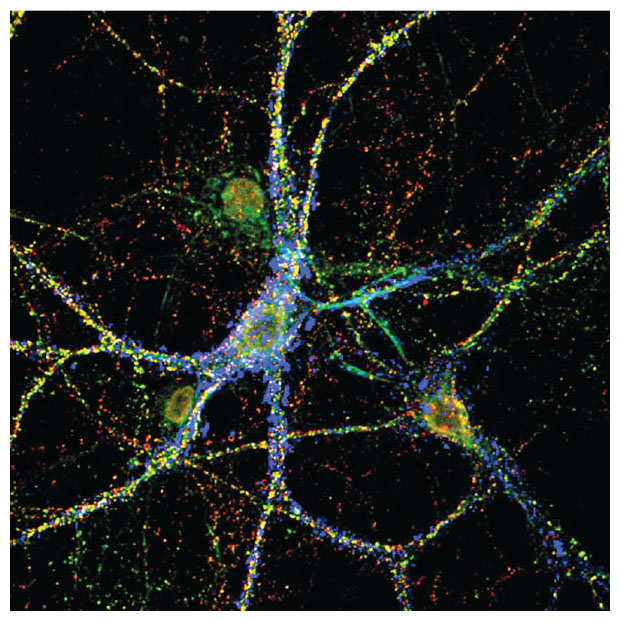
Sakina Palida, a graduate student in the lab of Nobel laureate Roger Tsien, explained how she and her colleagues had tackled what seemed to be a paradox: If, as is now known, most proteins in neuronal synapses undergo rapid turnover (within hours or days), how do the synapses themselves preserve memories over the course of a human lifetime? Reasoning that the basis for stability must lie outside the synapses but in close proximity to them, the researchers turned their attention to the perineuronal net. Working with cultured neurons and whole brain slices, they found evidence of this net enveloping mature neurons, where it restricted the formation of new synapses. Crucially, although the perineuronal net generally shows very little turnover, the research team found that it did erode under one condition: If an already-existing synapse became stronger as a result of exposure to a brain-stimulating chemical compound, the perineuronal net formed a hole at the site of the growing synapse.
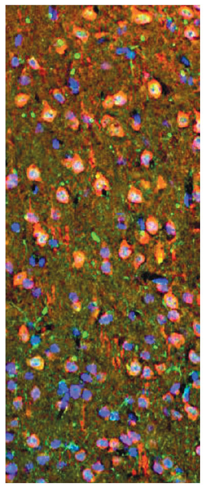
To test whether the pattern seen in the lab held true in live neurons, the Tsien team developed a technique for fluorescently labeling certain protein components of the perineuronal net in transgenic mice. When they saw the net undergo structural change, developing new holes in accord with the animals’ activity, the researchers knew they were on the right track. Further studies are eagerly anticipated to establish exactly which protein(s) are responsible for the durability of the perineuronal net and which ones account for its responsiveness to strengthening synapses.
Because the perineuronal net is so highly attuned to the state of nearby synapses, it's not surprising that this structural element also fills an important function in the developing brain. A recent paper by Janet Werker and Takao Hensch, of the Canadian Institute for Advanced Research, reviews the notion of so-called critical periods in development, when certain pathways in the brain have a heightened capacity to be shaped by experience; toward the end of this period, the perineuronal net acts as a “physical brake” to further synaptic growth. Conversely, the removal of perineuronal nets by injury or illness can re-open a window of opportunity for the growth of new synapses, to some extent enabling the nervous system to repair itself.
In the intimate and changeable relationship between the neurons and the multifunctional mesh that surrounds them, much remains to be explored. The next waning of interest in the perineuronal net may be a long time off.
Click "American Scientist" to access home page
American Scientist Comments and Discussion
To discuss our articles or comment on them, please share them and tag American Scientist on social media platforms. Here are links to our profiles on Twitter, Facebook, and LinkedIn.
If we re-share your post, we will moderate comments/discussion following our comments policy.