Paleontology's X-ray Excavations
By Catherine Clabby
Digital scans and computer-aided visualizations are liberating ancient life from stone.
Digital scans and computer-aided visualizations are liberating ancient life from stone.

DOI: 10.1511/2014.110.386
Paleontologists have always dedicated themselves to uncovering previously unseen aspects of life on Earth, and new imaging technology is taking them further toward that goal than ever before. Using high-resolution x-ray microtomography (micro-CT) they can look into both the exteriors and interiors of fossils at a microscopic scale, in three dimensions. (Tomography works by moving an x-ray source and detector around an object, building up an image layer by layer.) Best of all, the technique does not require slicing up the samples, so the same fossils can be examined again and again.
The resulting reconstructions have been a revelation to researchers such as Stephan Lautenschlager at the University of Bristol, who studies relationships between form and function in extinct vertebrates. He and his colleagues recently summarized the advances in a review article in Trends in Ecology & Evolution. Micro-CT and related imaging advances are “transforming our understanding of long-studied fossil groups, and of the narratives of organismal and ecological evolution that have been built upon them,” they write.
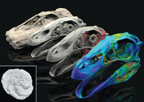
In one example, Lautenschlager has been studying the skull of a giant theropod dinosaur called Erlikosaurus andrewsi, which belonged to the same lineage as Tyrannosaurus rex but was, improbably, an herbivore with a beaked snout. When a scholar visiting from the University of Mongolia brought the E. andrewsi fossil to the United Kingdom, it was still partially embedded in stone. Lautenschlager and his collaborators performed a micro-CT scan to peer inside. Using the information from the digital view, the researchers virtually restored missing pieces of the cranial skeleton, beak, and jaw muscles, and reconstructed what the massive animal looked like when it was alive some 90 million years ago. Finally, the researchers performed an engineering test called finite element analysis on the digital model to examine its ability to withstand stress and strain. The results indicate that the dinosaur’s jaw wasn’t strong enough to chew tough leaves; more likely it swallowed them and processed the mass in its stomach. E. andrewsi’s beak probably protected the jaw from damaging strain.
Three-dimensional scanning is also proving valuable for learning more about ancient ecosystems from fossils of plants and invertebrate animals. Carole Gee, a paleobotanist at the University of Bonn, is exploring entire landscapes through her studies of small conifer seed cones. She turned her attention to the Upper Jurassic rocks of the Morrison Formation in the Western United States, strata that have yielded many dinosaur fossils but few plant fossils. The apparent scarcity of plants led some scientists to speculate that the region was mostly barren and semi-arid at the time. That conclusion didn’t make sense to Gee, because the large number of herbivorous dinosaurs living there would have needed an abundant food supply. So when she learned that private collectors had amassed 60 fossil conifer cones from the Morrison Formation, she set out to get some answers.
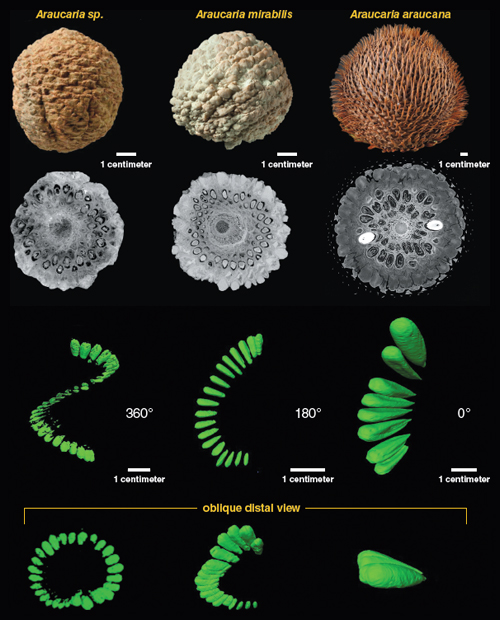
Using micro-CT, Gee was able to look into these rare fossils without cutting them into thin slices—a highly destructive, time-consuming process widely practiced in her field. She and her collaborators identified six cone types representing at least three different conifer families. Gee concludes that forests or woodlands were present and species-diverse in the Morrison Formation during the Late Jurassic. In addition, her scans open the door to detailed anatomical study of the seed cones, which could more precisely identify the conifer lineages of the Jurassic and place them in the context of plant evolution. “I can isolate different structures I want to see—ghostly structures—in 3D,” she says. “I can color-code each seed. I can look at the vascular system.”
Beyond the individual discoveries, Lautenschlager says that the rise of digital scanning technologies in paleontology will make it much easier for scientists to share data, addressing the limited access to fossil specimens that has plagued the field for so long. He and his Bristol colleagues call for a master archive that would house digital- study fossil data in standardized formats. They also argue that researchers should be obliged to share relevant primary tomographic data, along with any digital reconstructions, when they publish their research.
Gee disagrees about the mandatory sharing of data. Until recently, she notes, paleontologists went to the great time and effort to make thin slices of fossils and then they hung on to them for as long as it took to really study them in depth. At the same time, other scientists were always welcome to come by and have a look. Micro-CT scanning may be new, but the old standards should still apply, Gee says: “I want to keep my data until I figure it all out. If I have gone through all the effort to obtain the data, I would like to pull out all the information I need first, without any time pressure, before making the data set available to others.”
Click "American Scientist" to access home page
American Scientist Comments and Discussion
To discuss our articles or comment on them, please share them and tag American Scientist on social media platforms. Here are links to our profiles on Twitter, Facebook, and LinkedIn.
If we re-share your post, we will moderate comments/discussion following our comments policy.