Nanoscale Science
By Fenella Saunders
An interview with Paul S. Weiss
An interview with Paul S. Weiss

DOI: 10.1511/2025.113.1.7
Paul S. Weiss is a pioneering nanoscientist at the University of California, Los Angeles, where he previously directed the California NanoSystems Institute. He studies the ultimate limits of miniaturization, exploring the atomic-scale properties of surfaces, interfaces, and biomolecular assemblies. He has developed and applied atomic-resolution scanning tunneling microscopes and spectroscopic imaging methods to measure the structure, function, and spectra of the smallest switches and motors in the world. To do so, he and his group also developed chemical patterning methods to place molecules and to control intermolecular interactions from the subangstrom to centimeter scales. He applies these advances in many areas, including quantum information, sensing, neuroscience, microbiome studies, tissue engineering, cellular therapies, and high-throughput gene editing. Weiss was the recipient of the William Procter Prize for Scientific Achievement at the 2024 International Forum on Research Excellence (IFoRE), and spoke with editor-in-chief Fenella Saunders after the conference about his work. (This interview has been edited for length and clarity.)
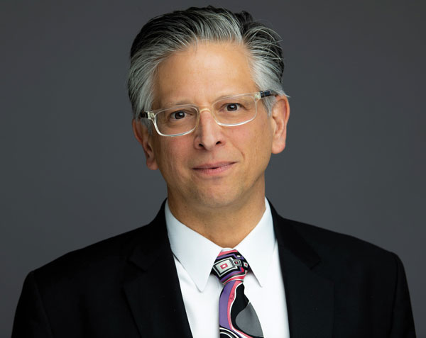
Courtesy of Paul S. Weiss
How did you end up specializing in nanoscience?
When I was an undergraduate, I got interested in how electronic structure and chemistry were coupled. I worked in crossed molecular beams of excited atoms, where we could point an orbital in space and see how that reacted. But I was looking for a more general way to approach the problem. I thought that on semiconductor surfaces, we could manipulate the occupation of the electronic orbitals. That would be a way to vary the chemistry.
The closest thing I could come up with was a group at Bell Laboratories, where people had figured out how semiconductors worked and invented the transistor. I worked with Mark Cardillo there, who had been slamming rare gas atoms into surfaces to excite the electrons on the semiconductor surface. I convinced him to start putting molecules on the surface, and we realized that we could detect a tiny fraction, a part in 100 million, of the reaction on the surface covered with the molecules we put down. What we didn’t know was where the molecules were on the surface and what they were doing chemically.
Right about at that time, during my PhD, the scanning tunneling microscope (STM) was invented, and really opened up the nanoscale world. We could start to image atoms. There was another postdoc at Bell Labs named Don Eigler who finished his time at Bell and moved to IBM Almaden in California. I followed him out there, and we built this low-temperature STM with the idea that we could not only measure the position of molecules, but we could do vibrational spectroscopy for chemical identification. We had this grand vision for which you needed an ultrastable microscope. This instrument was on its own foundation in a separate building inside the basement of the IBM building. Eventually, we made that experiment work. It took about 13 years. But it turned out to be not that useful in the end, because we don’t know the selection rules. Sometimes the signal goes up and sometimes it goes down. What we got out of that instrument that we built with this exceptional stability, where we could stay over a single atom for 10 days at a time, was that we could map the surface around an atom or molecule.
It turned out that Don and I both have a favorite rare gas: xenon. He had a dog named Xenon at the time. We put xenon down on the surface, just to see if we could image it. It didn’t have any electronic states near where we were probing. But sure enough, we could image those atoms. They were sitting out in the middle of an atomically flat terrace, which didn’t make any sense chemically. We reprogrammed the microscope so we could move the atoms out of the way to find out what was underneath. That was the first instance of moving atoms around with an STM and really showing that this microscope could do more than measure the structure. Later, Don spelled out “IBM” with atoms.
We could look at an isolated molecule that wasn’t moving on the surface, because it was at a very low temperature. We discovered that several atoms away, the electrons of the surface were perturbed. Normally, when we think about chemistry, a tiny change, a tenth of an angstrom, is enough to change from a single bond to a double bond. This was at 100 times greater distances. We could see with the microscope what turned out to be chemical effects. We looked at what roles those perturbations had, and it turns out they’re relevant in catalysis and building surface structures and a number of other areas.
It turns out there are other things we could do with the microscope as well. Spectroscopies let us look at all kinds of aspects of assemblies of molecules. Later, we got into the switches and motors on the surface, looking at the function in addition to structure and spectra. Now, we combine all those modalities together to do things like atomically resolve structures of the amyloid plaques that are thought to be responsible for neurogenic disease.
How did you create nanoscale switches?
There had been a discussion of whether a molecule could function as a wire, and later whether it could function as a conducting switch. We developed the means to isolate a single molecule in a controlled chemical environment, and then we could position our STM tip over it, and we could see it switching stochastically, and later we learned to drive it from one state to another. We developed two capabilities that turned out to be important. One is that we added the chemical dimension to nanolithography, controlling the exposed functional group on a surface. And then we also developed microscopes where we could sit our probe tip over the functional part of the surface, and then do the same measurement over and over, tens or hundreds or thousands of times, so we could work out what the mechanism of function is. In the first switches we looked at, there had been six different mechanisms proposed for how they worked. We systematically showed that all six ideas were wrong. We had to come up with our own mechanism, which turned out to be, again, chemistry related. When the molecules tilted, they changed the bonding to the surface, and that was responsible for the change in conductance.
How did you move from nanoscale molecular switches to motors?
One thing that’s fascinated me is how nature uses motors that convert chemical fuel to motion with more than 99 percent efficiency. My late colleague Paul Boyer figured out how those proton pumps in cell membranes work. They’re amazing. You take one ATP molecule as a fuel, you rotate the motor 120 degrees, and in three rotations, you pop a proton across the membrane. They’re so efficient that you can push the proton back through and get your fuel back. Nothing humans make at any scale is remotely that efficient. We use 50 to 75 kilograms of ATP every day, but we’re not made up of half chemical fuels. Rather, we can go back and forth between the fuel and its product many times every day in all of our cells.
At the same time we were studying these motors, I had a neuroscience colleague, Anne Andrews, come into my laboratory a little frustrated, because when she tried to capture proteins involved in neurotransmission, all kinds of biomolecules stuck to the surfaces. She looked at our control of the exposed chemistry on the surface and said, “Some of what you’re doing might be useful.” It was both flattering and insulting at the same time. I asked her what she meant and she said, look, the brain’s been doing nanoscience for hundreds of millions of years—you people are way behind. Let’s make a surface that prevents molecules from sticking and put, on average, neurotransmitters every five nanometers. We started working together. Later, we got married. We moved to UCLA together. That work led us into understanding that controlling the exposed chemistry at the nanoscale let us measure and control interactive biological systems.
Is that how you started working with the BRAIN Initiative?
The BRAIN [Brain Research through Advancing Innovative Neurotechnologies] Initiative came about because we were asked to put a panel together for the Office of Science and Technology Policy under the Obama White House, to come up with a grand vision of what would capture the public imagination about nanoscience. The part that I promoted really came from Anne’s work to listen in on the chemical communication in neural circuits and to understand how those circuits functioned, to be able to stimulate, predict, and understand the difference between healthy and diseased brains. We put together both the technologies for voltage measurements in parallel with chemical measurements. Anne leads that technology development for making chemically sensitive and specific arrays to go in the brains of animals, and soon also humans in some studies that are coming up.
This nanoscience group was brought back again to help develop the U.S. Microbiome Initiative in 2016. Some of the same sensors apply, but we needed oceanography and soil science and gynecology and dermatology and the gut microbiome. A lot of those technologies also work for wearable sensors. Anne has sensors developed for measuring cortisol in sweat as a continuous stress monitor, and one for continuous glucose monitoring.
How does the U.S. Microbiome Initiative connect to nanoscience?
The same sensors can target just about any biomarker. For example, in our mouths we have 200 or 300 different microbial species. Some are mutually exclusive. There’s one that gives you cavities and another one that doesn’t. You can have only one at a time in a particular place. One of our colleagues figured out what molecules they use to communicate. We can develop the tools to measure those and learn about how to avoid cavities just by having the right bacteria in our mouths.
We live with a kilogram or two of bacteria, some of which help us live and some of which are trying to kill us. You want to support the good ones and not the bad ones. We can do that with fairly simple choices of what we do day to day. But it’s quite amazing compared with all the treatments a person needs after they’re already in trouble.
How can nanoscience improve other disease treatments?
Steve Jonas, an MD–PhD student and resident in pediatric hematology oncology, worked out a project with us to come up with a way to do efficient, economical, safe, high-throughput gene editing to treat diseases such as sickle cell and thalassemia, in which there are 300,000 patients per year for each disease. If you replace 10 or 20 percent of the bone marrow with corrected cells, that’s a curative treatment. There are approved drugs for this replacement, but those are over $2 million a dose, so that doesn’t scale. We wanted to be able to do this treatment in one hour for a 12 kilogram child, which meant we needed to transfect a billion hematopoietic stem cells. And we wanted to be able to do it at many doctors’ offices around the world. With Steve and four other clinicians who do bone marrow transplants, we developed different ways to do this work. The same technology works for cancer immune therapy, for developing genetically modified T-cells.
Hsian-Rong Tseng in molecular pharmacology at UCLA had developed this sort of silica barbed wire to capture circulating tumor cells. He noticed that there’s penetration of these needles into the cells. He used host-guest chemistry to build carriers to get DNA, RNA, and protein machinery into the cell nucleus. But what he found with the barbed wire was, he could get those payloads into the cells, but just like if you jumped onto barbed wire, it’s easier to get on than it is to get off. He couldn’t get the cells off, but he could show that he could get those payloads into the cells.
“Nature uses motors that convert chemical fuel to motion with more than 99 percent efficiency. Nothing that humans make at any scale is even remotely that efficient.”
Before I came to UCLA, we’d studied what happens when you mechanically perturb membranes. We made what are called giant unilamellar vesicles. They’re like soap bubbles that mimic cells. We made a cup shape to mimic red blood cells, and when compressed, we noticed that pores opened up, to our surprise. We figured out how big the pores were and how long they lasted, but we had no idea what to do with that. Later, Robert Langer, Dan Anderson, and Klavs Jensen at MIT showed that if you pushed cells through a constriction in a microfluidic channel, you opened up transient pores, as we had in the model cells. You could deliver payloads to the cell nucleus that way. So we realized, that’s what you do with it. The struggle they had was their channels would clog. We instead used acoustics to define a virtual channel. It turns out one of our UCLA alumni, Tony Huang, had moved to Duke University. We called him up and said, let’s define virtual channels in this way and see how effective that will be. I sent four students, and it took them a week to make the whole thing work. The nice part about that is, there’s no constriction. The channels cannot clog. Every time you change cell types, you need to optimize the diameter and so forth, and with acoustics all you do is change the waveform. One device will serve all.
I assumed it would be best to make the constriction down the center of the channel, but we found out that it works best to use acoustofluidics to charge the outside of the payloads to hold them on one wall, and then bounce the cells off the wall using the acoustics. We can do 12 million cells per channel per hour at peak efficiency. We made that parallel, so with 100 parallel channels, that’s more than a billion cells. Cell viability is very high, more than 90 percent. Right now, the efficiency is in the 20 to 40 percent range, depending on the particular cell, which is sufficient. It’s very exciting that we can do this. We haven’t tested it in animals or humans yet at all, but that’s coming up shortly.
How else can you use nanoscience to manipulate cells for other purposes?
A PhD student, Amir Nasajpour, got an idea from cellular agriculture, trying to grow meat and fish in a laboratory, in which there are difficulties with the scaffold. It’s often gelatin, which comes from boiled cow bones, which kind of defeats the purpose of not killing animals to grow meat in the lab. Or there are scaffolds that are plastic or silk, but they need to be decorated with proteins that again come from animal sources. In addition, it’s hard to scale the growth, because you’re trying to grow muscle, so you need to stimulate it either mechanically or electrically. In addition, we don’t just eat muscle. We eat muscle and fat in particular arrangements, depending on preferences. It’s difficult to co-culture in the current setups.
Amir reverse engineered all that and came up with a combination of molecules that come from plants that have been known since the 19th century and form a liquid crystal. All the relevant cells recognize them, so the adhesion is built into the system. They’re also FDA-approved. They’re in cosmetics, so they’re produced at scale. He sets the formulation of these molecules so their transition temperature is the incubator temperature. It turns out that bioreactors are not perfect in temperature control. They’re not made to be. The small fluctuations in temperature give rise to these very large motions of the underlying scaffold, which stimulates the cells. And all the cells recognize those scaffold molecules, so we don’t need to encapsulate them.
Then we wanted to get a 3D movie of the growth. We went to our superresolution microscopy facility and gave them the scaffold to grow any cell of their choice. A few hours later, we got a call saying, sorry, we used this eye cancer, uveal melanoma, cell line that we like to use as a model, and the organoid grew too quickly for us to measure. We just looked at the phone and said, what? We went through a whole bunch of other cancer cell lines. We’ve now grown lung cancer, colon cancer, and pediatric brain cancer in a cell line nobody else could grow. We can also co-culture, because to make a biological twin of a tumor, you need to have the healthy cells around to recapitulate the tumor microenvironment. We’ve integrated with the robotic high-throughput screening that we have, the same as a drug company would use for personalized medicine, for individual patients, so you can test potential therapeutics against many copies of a tumor. And then you can also use that to look at several different patient lines and test potential therapeutics. If we can do it on the time scale of treatment, then we’re in good shape to go ahead.
Click "American Scientist" to access home page
American Scientist Comments and Discussion
To discuss our articles or comment on them, please share them and tag American Scientist on social media platforms. Here are links to our profiles on Twitter, Facebook, and LinkedIn.
If we re-share your post, we will moderate comments/discussion following our comments policy.