Mapping Macromolecules in Cells
By Catherine Clabby
Advanced biophotonics helps researchers observe the inner workings of living cells
Advanced biophotonics helps researchers observe the inner workings of living cells
DOI: 10.1511/2011.88.66
Researchers at the State University of New York at Buffalo are using advanced biophotonics to observe multiple types of macromolecules at once in living cells. The Institute for Lasers, Photonics and Biophotonics, directed by Paras N. Prasad, was first to apply this technology to monitor the distribution of native proteins, lipids, RNA and DNA during apoptosis, or programmed cell death. The work was published in 2010 in the Proceedings of the National Academy of Sciences of the U.S.A. More applications are in development. Artem Pliss, an institute research assistant professor, described the method and its promise to American Scientist associate editor Catherine Clabby.
A.S. What are the technologies you used?
A.P. Aliaksandr Kachynski and Andrey Kuzmin here in Buffalo designed and operate our multiplex setup. With it, we integrate two advanced nonlinear optical modalities: coherent anti-Stokes Raman scattering (CARS) and two-photon excited fluorescence (TPEF). CARS records the vibration frequencies of molecular bonds after they are exposed to intense bursts of light. We can use CARS to image protein and lipid macromolecules in cells, but the RNA and DNA vibration frequencies are barely distinguishable. So we selectively stained RNA and genomic DNA with fluorescent dye and used TPEF to image those macromolecules in two different fluorescent channels. The combination of technologies permits the characterization and monitoring of intracellular molecular organization at a level of sensitivity and complexity that previously was unachievable.
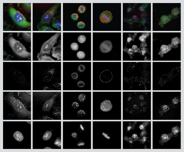
Images courtesy of the Institute for Lasers, Photonics and Biophotonics at the State University of New York at Buffalo.
A.S. How does this contribute to the practice of cell imaging?
A.P. Traditionally, cellular organization is studied by the imaging of particular macromolecules tagged by fluorescent markers, which captures their distribution and interactions. This conventional technique, however, omits the locations of millions of unstained macromolecules, inherently limiting our understanding of cellular organization. In our study, we developed this hybrid approach, in which the specific molecules are mapped by TPEF simultaneously with the mapping of the cell’s proteins by CARS. The combination provides insights into the dynamic transformations of the cellular structure. In the apoptosis study, we observed proteins change from being distributed nearly uniformly in the cell nucleus to being present in progressively irregular patterns, a transition not described before this study.
A.S. Can you give an example of how this will be useful?
A.P. This can be used to observe different organelles that accommodate specific biochemical activities. Revealing the macromolecular patterns in these structures can serve as a real-time indicator of cellular physiological condition. Our approach will also be valuable for monitoring the subcellular configuration of macromolecules during drug-cell interactions, which could facilitate drug development.
A.S. What improvements are coming?
A.P. We are working on expanding our imaging by adding several other optical modalities. Our aim is to simultaneously acquire diverse layers of information, such as the distribution, interactions, local concentrations and interface properties of different types of macromolecules. That will give more comprehensive characterization of the molecular structure in cells and tissues. We anticipate that our technology will be used for noninvasive molecular diagnostics as well as for drug development, where it will allow researchers to collect specific information on intracellular structure and enable high-throughput drug screening at the level of individual cells.
In Sightings, American Scientist publishes examples of innovative scientific imaging from diverse research fields.
Click "American Scientist" to access home page
American Scientist Comments and Discussion
To discuss our articles or comment on them, please share them and tag American Scientist on social media platforms. Here are links to our profiles on Twitter, Facebook, and LinkedIn.
If we re-share your post, we will moderate comments/discussion following our comments policy.