3D Carnivorous Plants
By Catherine Clabby
Optical projection tomography aids studies of predatory plants.
Optical projection tomography aids studies of predatory plants.

DOI: 10.1511/2013.100.56
Prominent molecular biologist Enrico Coen has discovered multiple classes of regulatory genes that are vital to the evolution and development of flowers. His research team—based at the John Innes Centre in England—is also investigating genes that produce plant leaves of specific sizes and shapes. To that end, Karen Lee, a researcher in Coen’s lab, is ramping up studies of carnivorous plants. Lee uses optical projection tomography (OPT), a relatively new type of microscopy, to get a better look at their growth and shape. Created to study gene expression in mouse embryos, OPT provides three-dimensional views of developing plants unavailable from any other technology, Lee told American Scientist contributing editor Catherine Clabby.
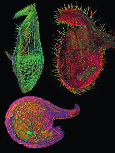
Optical projection tomography images are courtesy of Karen Lee, Jerome Avondo, Grant Calder, Andrew Bangham and Enrico Coen at the John Innes Centre..
Why are you interested in carnivorous plants?
These plants may help answer the question of how the vast variety of leaf and flower forms construct themselves from small groups of cells. Our team uses a combination of imaging, mathematical modeling and genetic approaches to determine the rules by which genes control the shaping of leaves and flowers. This approach helps us understand mechanisms of leaf growth in the model plant species Arabidopsis thaliana, which has simple leaves. Carnivorous plants are fascinating because their leaves display complex shapes that allow them to entice, capture and devour animal prey. We would like to know how these shapes are generated and how they evolved from species with simpler shapes.
Can such unusual plants be good model organisms?
Bladderworts (Utricularia), Monkey cup pitcher plants (Nepenthes), the Albany pitcher plant (Cephalotus) and other pitcher plant genera (such as Sarracenia and Darlingtonia) all independently evolved cup-shaped leaves with lids. Common developmental rules may underlie these different forms. To get at the key developmental mechanisms, we need a model carnivorous plant that is easy to grow, image and genetically dissect. The bladderwort Utricularia gibba is perfect. It has a tiny genome, which makes it amenable to sequencing and gene discovery.
Why is it hard to observe plant growth and development?
It is challenging to capture how cells and tissues grow in three dimensions (3D) and to quantify changing shapes through development. A plant must be kept alive and growing while being imaged over many days. We use a combination of confocal and OPT microscopy, together with image analysis tools, to study plant development. Confocal microscopy captures growth of the molecular skeleton within cells and tracks cell growth and division in developing primordial structures. But because it focuses a laser beam to a fine point, it cannot be used on samples larger than 1 millimeter. OPT works by projecting light through a specimen at multiple angles. A detector captures the shadows produced as the specimen is rotated. Computer software then assembles 3D images of full plant structures from the information obtained with the detectors.
What is the value of 3D imaging for this research?
For anything other than a growing linear file of cells or a flat sheet of expanding tissue, growth involves deformations in three dimensions. OPT allows us to compare and measure such deformations as a complex leaf or flower develops and grows. With OPT we can also see inside a specimen. And differences between mutants and normal leaves or flowers can be visualized more clearly using 3D images. Gene expression patterns, visible with markers, are often looked at in a slice of tissue on a microscope slide. But it can be difficult to know where this slice came from in a plant. OPT overcomes this problem and allows us to see where a gene is acting in the context of a whole organ, a functional or structural unit in a plant. In carnivorous plants, that can include bladders, the cup-like leaf structures that trap prey.
Will you combine gene expression and imaging studies?
We plan to screen for Utricularia bladder mutations and use OPT to compare mutant bladders with normal ones. This will give us an idea of the role played by the gene that was inactivated in the mutant. We intend to sequence the genome of the mutant bladder and compare the sequence to the normal bladder genome to find the gene we have knocked out. Once we have the sequence of the mutant form of the gene, we can use it as a probe to look at where the gene is expressed in slices of normal bladder tissue on microscope slides. OPT of whole bladders could allow these slices to be mapped to the whole bladder structure. This will give us clues to the role the gene plays in bladder developmental regulatory networks. Information obtained in this way will help us understand how genes control bladder growth and how they differ from genes controlling growth of a simpler plant leaf. If we find simple rules underlying complex leaf formation in carnivorous plants, we may find the general principles by which complex forms in both plants and animals arise.
Click "American Scientist" to access home page

American Scientist Comments and Discussion
To discuss our articles or comment on them, please share them and tag American Scientist on social media platforms. Here are links to our profiles on Twitter, Facebook, and LinkedIn.
If we re-share your post, we will moderate comments/discussion following our comments policy.